Radiation imaging and detection group
We develop new solid state materials for the detection of X-rays, gamma rays and neutrons, and for recording images of objects placed in X-ray or neutron beams.
Our aim is to design and make imaging and detection materials with superior performance—greater sensitivity and speed for detector materials, and enhanced contrast and resolution for imaging—and to demonstrate their performance by developing practical dose-measurement and imaging systems for commercial uptake.
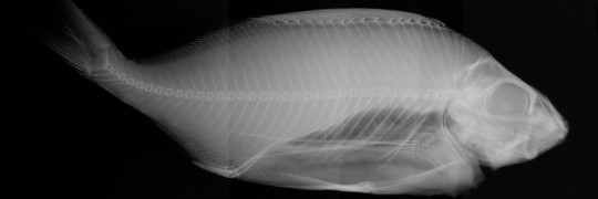
Imaging
For imaging, we develop ceramic, glass ceramic, and composite ‘storage phosphors', where a latent image is stored in the material until it is read out by a laser. The image appears as light-induced phosphorescence wherever the X-rays were strongest.
We also work on prompt phosphors, where the phosphorescent emission occurs at the same time as the X-ray irradiation.
Detection
For the detection of ionizing radiation, we focus on inorganic ceramic materials and composite materials that exhibit radioluminescence and optically stimulated luminescence. The dose rate can be determined during irradiation by radioluminescence where ionizing radiation results in the emission of visible light.
The dose can be obtained after irradiation from optically stimulated luminescence where ionizing radiation creates trapped electrons and holes that can be stimulated with light leading to the emission of light at another wavelength.
Applications
Applications for this technology are in radiation therapy, radiography for medical, veterinary and dental practice, non-destructive testing, and security applications. We are developing products that use our dosimetric and imaging materials.
Research team
Our group comprises physicists, chemists, and electrical engineers based at Victoria University of Wellington, Wellington Hospital, Callaghan Innovation, i2M Labs, and GNS Science. Read more about our current research projects.