Product development
Technology overview
We are developing technology for optics-based (1) X-ray imaging and (2) radiation dosimetry. The main methods are radioluminescence (RL) for real-time imaging and dosimetry and optically stimulated luminescence (OSL) for X-ray storage phosphors and cumulative radiation dose monitoring.
The physics of radioluminescence involves ionising radiation (for example, X-rays, gamma-rays, neutrons) causing the excitation of electrons and holes in the detector. They then combine at a luminescence ion and result in light that is detected.
In OSL the electrons and holes become trapped at carrier traps. At a later time, they can be optically stimulated out of the carrier traps by illumination of light at one wavelength and then recombine at a luminescence ion and produce emitted light at another wavelength. The intensity of the emitted light can be related to the ionising radiation dose and used to contract a 2D dosimeter image.
X-ray storage phosphor research and technology
The potential applications include quality assurance for medical (for example, X-ray imaging for emergency services, research, replacing conventional film X-ray images, dose verification and validation for radiation therapy), non-destructive testing (for example, detection of cracks in metal and plastic structures), border security (e.g. imaging of packages and containers), veterinary (for example, on-site X-ray imaging of animals), water content monitoring (for example, soil moisture, timber water content, etc.), energy (for example, coal/ash content), and agricultural (for example, imaging of apples etc.).
We are researching glass-ceramics (micron sized crystallites in a glass matrix) and crystallite/polymer mixes. The glass-ceramic research is covered by our US patent, and has been licensed to Materials Development Inc.
The medical and nondestructive testing imaging markets are dominated by indirect imaging via x-ray storage phosphors and direct imaging (for example, via a-Se detectors from Siemens). However standard radiation imaging plates from Siemens, GE and AGFA have moderate pixel sizes (to 0.07 mm) but the spatial resolution is >0.1mm. We have shown that a spatial resolution of at least 0.01mm is possible with our materials.
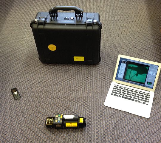
We have developed a cost-effective portable X-ray read-out unit for digital and computed radiography. The system is highly portable, and is controlled either by a smartphone/pad via wireless or a laptop computer via USB links. The potential applications include non-destructive testing, farm vets, and emergency services.
This system works extremely well with existing commercial imaging plates for non-destructive testing and security applications, and we have recently found that it also shows acceptable images with veterinary and medical doses

Video of toy cockroach
Dosimeter research and technology
Radiation dosimeters are used to monitor and detect ionising radiation doses and dose rates. In our case we are focusing on RL and OSL dosimeters
We have been researching bulk and nanoparticle fluoride compounds and we have a USA patent on key fluoroperovskite dosimeter materials and a point dosimeter patent. The advantages of the materials that we are researching include transparency, near tissue equivalence, and a high saturation dose.
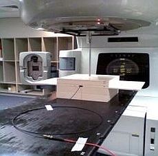
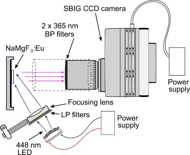
The material’s response to radiation has been tested with a medical linear accelerator operating at 6 MV (right figure) and shown to have an acceptable energy independence.
We are focusing on applications that require dosimeters that respond to radiation in manner that is similar to tissue. One key application area is dose verification and validation for the radiotherapy treatment of cancer and especially when medical linear accelerators are used. This has recently become more important with the introduction of volumetric modulated arc radiotherapy.
We are developing 2D dosimeter prototypes and testing two different concepts.
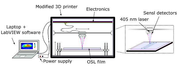
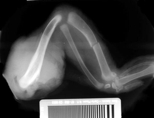
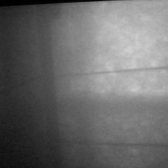
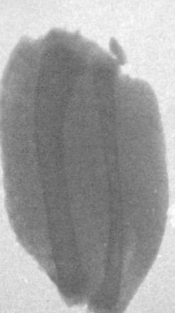
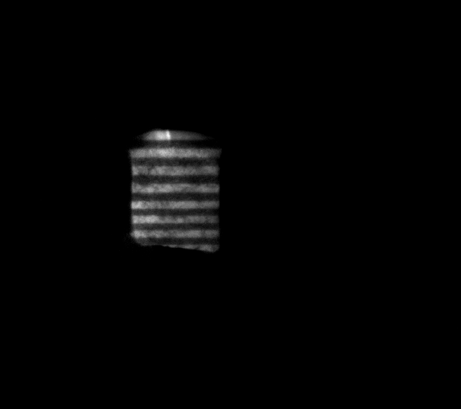
The second system uses a high-quality camera to rapidly take the 2D dose image in as little as 30 seconds (right figure). We have already shown that it can take a low dose image after X-ray irradiation. This can be seen in the figure below, which is an X-ray image of an X-ray phantom
Patents
Christian Josef Dotzler, Andrew Edgar and Grant Victor McLelland Williams, "Fluoroperovskite radiation dosimeters and storage phosphors", US2010/0200741 A1.
Grant Williams, "Radiation dosimeter detection system and methods", Patent number EP2616844
Peter Willems, Johann-Martin Spaeth, Stefan Schweizer, Andrew Edgar, Luc Struye and Paul Leblans, "Fluoro glass ceramic showing photostimulable properties", United States Patent No. US 6352949 B1, 5 March 2002