Current research
Optics-based X-ray imaging systems and high-resolution imaging plates
Andy Edgar
There has been a revolution in X-ray imaging in the last twenty years, with traditional photographic film/phosphor methods being replaced by digital techniques, including storage phosphor, CCD/phosphor, CMOS/phosphor and photoconductor array methods.
Storage phosphors based on the optically stimulated luminescence effect offer many advantages over film but suffer from inferior spatial resolution. The problem lies in the light scattering from the powder grains of the storage phosphor (usually BaFBr:Eu). This problem rules out storage phosphors for mass radiography applications such as mammography.
Our research into nano-structured materials such as glass-ceramics and polymer-nanocrystal composites seeks to combine good storage phosphor properties with high transparency for computed and direct X-ray radiography.
Radiation Dosimeters
Grant Williams
We are researching radiation dosimeter with an emphasis on optics-based materials and devices. Our focus is on materials that have a response to radiation that is similar to tissue where the applications include quality assurance for medical linear accelerators and other radiation sources.
We are studying bulk and nanoparticle fluoroperovskites where the materials exhibit radioluminescence during exposure to ionizing radiation and optically stimulated luminescence after exposure to ionizing radiation.
We have shown that the optically stimulated luminescence is linear up to high doses and the materials can be processed so that the real time signal is independent of the dose history.
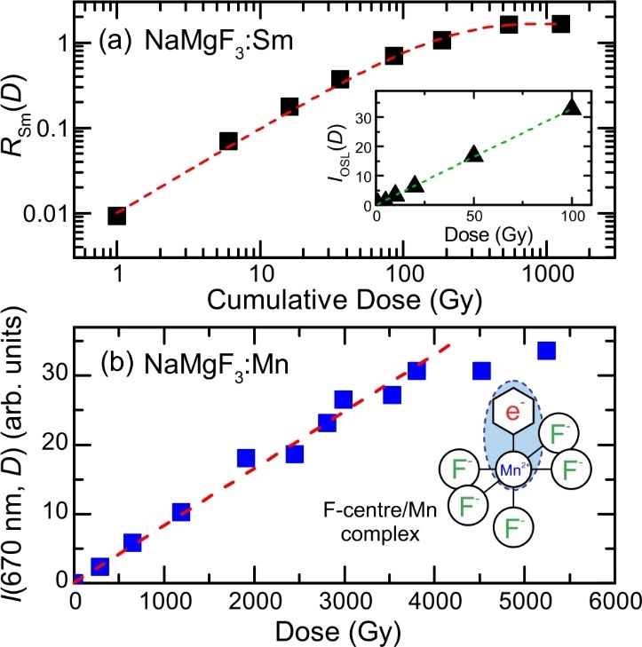
As an example we show in the upper figure that the optically stimulated luminescence response from NaMgF3:Sm in linear up to high doses. We have also found an optically active point defect that arises from an F-centre/Mn complex. The signal from this is linear to very high doses (lower figure).
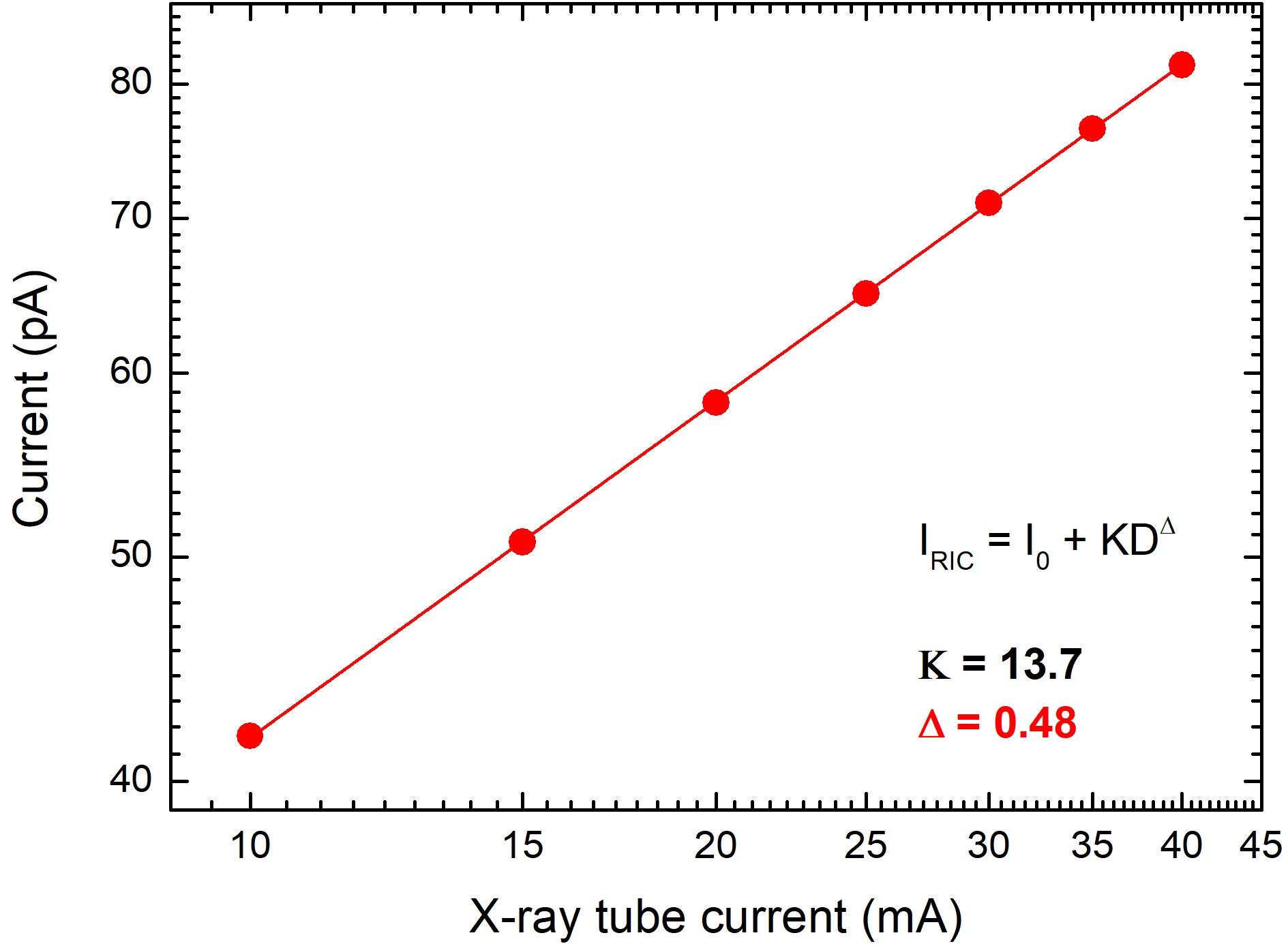
We have recently shown that NaMgF3 doped with a luminescent ion displays a current that is directly related to the X-ray dose rate. This provides for a novel tissue-equivalent dosimeter where the dose during irradiation can be monitored by electrical current and radioluminescence to enhance quality assurance.
Related resources
Radiation instrumentation and consulting
X-ray imaging resources
- Daresbury Imaging Group
- University of Leeds
- Micro Photonics.
Scintillator resources
Dosimetry resources
- A Practical Course in Reference Dosimetry
- Dosimetry for X-Ray Users
- NIST Radiation Interactions and Dosimetry Group.
Patent databases
- Online searchable patent and trademark database: United States Patent and Trademark Office
Online searchable database of New Zealand patents: Intellectual Property Office of New Zealand.